ads/wkwkland.txt
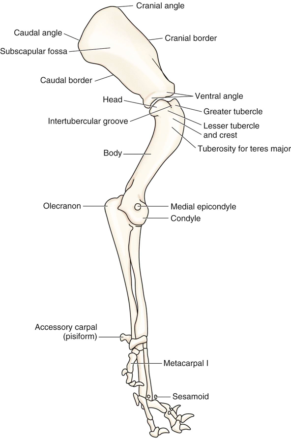
36 Top Pictures Cat Hindlimb Bone Anatomy - How Do Cat Paws Work How It Works. The anatomy of the domestic cat is similar to that of other members of the genus felis. The most common cause of amputations in cats is due to severe trauma, usually as a result of a traffic accident. More fragile bone of the lower hindlimb. Learn anatomy cat hindlimb with free interactive flashcards. It is made up of skeletal bones, muscles, cartilage, tendons, ligaments, joints and connective tissue.
ads/bitcoin1.txt
Learn vocabulary, terms, and more with flashcards, games, and other study tools. This allows freedom of movement of the foreleg, which can be turned in almost any direction. The ligaments in a cat are a tough band that is composed of a white, slightly elastic, fibrous tissue that binds the ends of bones together. Wilson1 1structure and motion laboratory, the royal veterinary college, university of london, hawkshead lane, north mymms, hatfield, hertfordshire, uk 2research department of the national zoological gardens of south africa. Common orthopaedic conditions in cats include forelimb fractures and femoral (large hindlimb bone) fractures.
Osteosarcomas and fibrosarcomas are two cancers which can develop in the leg.
ads/bitcoin2.txt
The pubis and ischium bones have an obturator foramen (hole). The musculoskeletal system is responsible for form, support, stability and movement. Diagram of the skeleton of a cat. Learn vocabulary, terms, and more with flashcards, games, and other study tools. Functional anatomy of the cheetah (acinonyx jubatus) hindlimb penny e. The anatomy of the domestic cat is similar to that of other members of the genus felis. Ch 7 anat 95 terms. Arrows b & d point to the papillae used for taste. It is an inverted 'u' shape from which the femur bone forms the hip joint. The cat bone structure also provides a framework around the vital organs while manufacturing blood cells and stockpiling important minerals. Treatment is femoral head and neck excision or total hip replacement. The ligaments in a cat are a tough band that is composed of a white, slightly elastic, fibrous tissue that binds the ends of bones together. The anatomy of domestic animals, 5th edition, sisson and grossman, wb saunders co., philadelphia, 1975.
Clancy,1* emily lane2 and alan m. This allows freedom of movement of the foreleg, which can be turned in almost any direction. It is the largest of the carpal bones (it is the end result of the fusion of the radial, central, and intermediate carpal bones), and it articulates proximally with the radius, laterally with the ulnar carpal bone, and distally with all four of the. The ligaments in a cat are a tough band that is composed of a white, slightly elastic, fibrous tissue that binds the ends of bones together. Main muscles of the trunk.
From a visual inspection of these plots, we can observe that although most sabretooths plot within the morphological range of conical‐tooth cats, some of them have extremely robust fore‐ and hindlimb bones.
ads/bitcoin2.txt
Diagram of the skeleton of a cat. The obturator nerve and artery pass through this hole to supply blood and control some of the muscles of the hindlimb. A cats skeleton is very similar to that of a human being, however it does lack the shoulder blade bones. It is the largest of the carpal bones (it is the end result of the fusion of the radial, central, and intermediate carpal bones), and it articulates proximally with the radius, laterally with the ulnar carpal bone, and distally with all four of the. Radiographs show a slipped femoral epiphysis, there may be 'apple coring' of the femoral neck. Wilson1 1structure and motion laboratory, the royal veterinary college, university of london, hawkshead lane, north mymms, hatfield, hertfordshire, uk 2research department of the national zoological gardens of south africa. The three components of each hip bone are the ilium, pubis and ischium. Some cancers can affect the leg including bone cancer and vas (vaccine associated cancer).this is a rare cancer with an incidence of 1 in 1,000 to 1 in 10,000 vaccinated cats. More fragile bone of the lower hindlimb. The pelvic girdle is formed by two hip bones which are joined ventrally at the cartilagenous pelvic symphysis and articulate dorsally with the sacrum. See all 9 sets in this study guide. All cats are capable of walking very. Other bones include the jawbones (mandible and maxilla), nasal bones, cheekbones and eye orbit.
All cats are capable of walking very. The obturator nerve and artery pass through this hole to supply blood and control some of the muscles of the hindlimb. Arrows b & d point to the papillae used for taste. Some cancers can affect the leg including bone cancer and vas (vaccine associated cancer).this is a rare cancer with an incidence of 1 in 1,000 to 1 in 10,000 vaccinated cats. The anatomy of domestic animals, 5th edition, sisson and grossman, wb saunders co., philadelphia, 1975.

Bones of the skeleton 63 terms.
ads/bitcoin2.txt
The ilium makes up the craniodorsal part of the hip bone. In the pubis bone, is the acetabulum which is the hip socket. Bones of the skeleton 63 terms. The bone that articulates with the hip bones to form the hip joint is the femur. From a visual inspection of these plots, we can observe that although most sabretooths plot within the morphological range of conical‐tooth cats, some of them have extremely robust fore‐ and hindlimb bones. Main muscles of the trunk. Learn vocabulary, terms, and more with flashcards, games, and other study tools. You can use the diagram of the pigeon skeleton (figure 9.3) to find the names of the hind limb bones. The thick skull bone protects the cat's delicate brain; The cat bone structure also provides a framework around the vital organs while manufacturing blood cells and stockpiling important minerals. Very similar to ours but for some reason cats decided to walk and run on their toes or the end of the metacarpus. Start studying anatomy cat muscles. It is an inverted 'u' shape from which the femur bone forms the hip joint.
ads/bitcoin3.txt
ads/bitcoin4.txt
ads/bitcoin5.txt
ads/wkwkland.txt
0 Response to "36 Top Pictures Cat Hindlimb Bone Anatomy - How Do Cat Paws Work How It Works"
Posting Komentar